At Trinidad Vision Center, we recognize the vital role healthy eyes play in your daily life and overall well-being. Our goal is to identify and manage eye conditions early—before they lead to more serious complications or vision loss. Serving San Antonio and surrounding communities, our skilled team is dedicated to providing personalized, compassionate care that addresses both acute concerns and long-term prevention. Below, you’ll find details on some of the most common conditions we encounter.


A cataract occurs when the eye’s natural lens becomes clouded, resulting in blurry or dimmed vision that can impact daily tasks such as reading or driving. Cataracts often develop slowly, so early detection is critical. During routine exams, we use advanced diagnostic tools to identify cataracts in their initial stages. If needed, we can discuss surgical options that replace the clouded lens with a clear artificial lens, helping restore clarity and improve quality of life.

Diabetic Eye Disease refers to a group of vision-threatening conditions—most commonly diabetic retinopathy—that can develop in individuals with diabetes. High blood sugar levels can damage the fragile blood vessels in the retina, potentially leading to permanent vision loss if left untreated. At Trinidad Vision Center, we conduct detailed retinal evaluations and offer targeted management strategies, including monitoring, laser treatments, or other interventions, to help preserve your eyesight for years to come.

Glaucoma is often called the “silent thief of sight” because it can progress undetected until irreversible vision loss has occurred. This group of diseases is typically linked to increased eye pressure, which damages the optic nerve over time. We perform specialized tests to measure intraocular pressure and monitor optic nerve health, allowing us to catch glaucoma in its early stages. Treatment may include medicated eye drops, laser therapy, or minimally invasive procedures—all aimed at halting or slowing further damage.
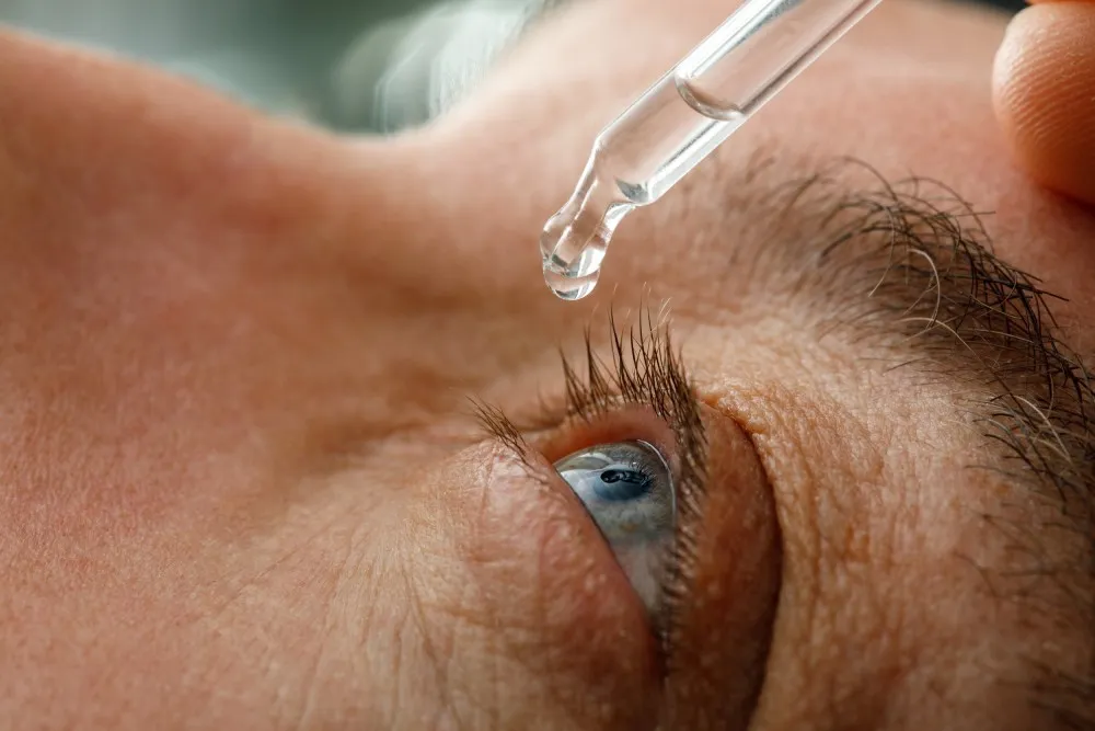
Dry Eye Syndrome arises when your eyes don’t produce enough tears or the tears you do produce are of poor quality. This can lead to chronic discomfort, redness, and fluctuating vision. Factors like environment, age, and medical conditions can all contribute to dry eye. Through comprehensive evaluations, we identify the specific cause of your dryness and develop a personalized plan—ranging from lubricating drops to in-office treatments—to provide relief and enhance tear quality.
If you’ve noticed any changes in your vision or suspect you may be experiencing one of these conditions, our team is here to help. We’ll begin with a comprehensive eye exam, discuss your concerns, and determine the most effective approach to preserve and optimize your eyesight.


Dr. Jake Trinidad is an esteemed ophthalmic surgeon with deep San Antonio roots, having graduated from Clark High School, Trinity University, and UIW before pursuing his medical degree at Indiana University School of Medicine and completing his ophthalmology residency. With over 10 years of experience and more than 10,000 successful eye surgeries, he has earned a trusted reputation for his precision, expertise, and compassionate approach—particularly in cataract surgery and advanced eye treatments.
Driven by his commitment to patient-centered care, Dr. Trinidad returned to his hometown to serve the community that shaped him, offering personalized, family-oriented services that ensure every patient feels truly valued. At Trinidad Vision, we see more than just your eyes—we see you. Experience the difference with trusted care, expert solutions, and a team dedicated to your well-being.


Your journey toward a healthier vision begins with a thorough evaluation at Trinidad Vision Center. Whether you’re experiencing specific symptoms or simply need a checkup, our team will conduct a detailed exam and listen closely to your concerns.
We leverage modern imaging and testing technology to detect a variety of conditions, from early-stage cataracts to diabetic changes in the retina. By identifying these issues promptly, we can create targeted solutions that safeguard your long-term vision.
After gathering all necessary diagnostic information, our specialists outline a personalized plan to manage or treat your condition. This could include medications, lifestyle modifications, specialized therapies, or surgical interventions—always tailored to your unique needs and goals.
Trinidad Vision Center’s San Antonio facility offers a comfortable, welcoming atmosphere that embodies our Texas Hill Country heritage. Patients often describe our cozy, thoughtfully designed interior as calming—setting the stage for an exceptional care experience. Our dedicated team strives to ensure you feel supported and informed at each visit, whether you’re here for a routine check or a more specialized procedure.


“This was the most comfortable Dr appointment I’ve ever had.. there was no waiting.. the machines for checking out your eyes were so easy.. and I was so happy to reunite with a first grade student that I had the pleasure of teaching .. Dr. Trinidad!! What a sweet surprise..
His office has great technicians and the atmosphere was very welcoming !! I’ve found the ophthalmologist that I was looking for !!"
“They are well organized. Office wait time has been very short, about 5 minutes each time I have visited! Just had surgery and now I have 20/20 vision. I highly recommend Dr Trinidad”
“I’m almost 86 years old been to a lot of ophthalmologists and he is without a doubt the most communicative, caring, brilliant, best kept secret in San Antonio! Highly recommend not doing anybody with him he has it all.”
"My visit with Dr.Trinidad and his staff was for my annual Diabetic eye exam. It went extremely well, Dr Trinidad's staff made me feel very much at ease. After Dr. Trinidad examined my eye's.He took his time to explain his findings and scheduled a followup.I would highly recommend Dr. Trinidad. He was very compassionate and professional and gave me peace of mind. Thank you so much to Dr. Trinidad and staff."
"I had an excellent experience with Dr. Trinidad and his medical assistants and other employees... The Dr. and his staff was wonderful, and they were able to diagnose my problems effectively, providing me with the information and advice needed."
"Dr Trinidad and his staff are a great team. They are well organized. Office wait time has been very short, about 5 minutes each time I have visited! Just had surgery and now I have 20/20 vision. I highly recommend Dr Trinidad"
"Absolutely the warmest Dr. Visit I had in awhile, the communication how Dr. Trinidad explained everything so detailed was very impressed. Honestly felt like I was talking to a childhood friend 👌. Nicole as a representative at the front was incredibly pleasant. They also had T. Ford, Versace, CHANEL and offer botox (I think)."
"Everyone had patience, was understanding, and went above and beyond to calm my nerves through out my whole visit. Not only did Dr.Trinidad take his time to make sure I understood what he was seeing/going on with my eyes but he truly does care and is passionate about his work. I definitely will be making him the person I go to for all my eye visits."

Many insurance plans do provide coverage for diagnosing and managing cataracts, glaucoma, and other eye conditions. The extent varies by plan, so our team will be happy to review your benefits and discuss any out-of-pocket costs.
For most healthy adults, an annual or biennial exam is sufficient. However, if you have a condition like diabetes or a family history of glaucoma, more frequent visits may be necessary to detect issues early.
If your condition progresses or you develop more severe symptoms, we’ll discuss additional treatment options. This might include medication adjustments, laser therapy, or surgical interventions, depending on your diagnosis and overall eye health.

Don’t wait until symptoms worsen—book your consultation today to safeguard your vision for the future. Our caring, knowledgeable staff stands ready to guide you from diagnosis through every stage of treatment, ensuring you receive the highest standard of ophthalmological care.


